Orbital Tumors - Dermoids

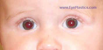
Pre operative photo: note the mass along the left, outer bro w

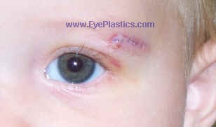
5 days after surgery, sutures in place
Etiology
- benign cystic lesions that are choristomas (tumors composed of tissues not usually found at the involved site)
- originate at bony suture sites during embryogenesis as a lead to of surface skin elements becoming entrapped
- usually found in early childhood (25% are noted at birth), but can also be diagnosed in adults
- in adults, they often involve the deep orbital tissues and grow to a very large size
- Not to be confused with Epidermoid cysts which are similar but lack certain elements in the wall of the cyst
Differential Diagnosis
- encephalocele
- lacrimal mass/tumor
- lacrimal mass or tumor
Work-up / Course / Prognosis
- CT/MRI imaging reveals characteristic round to oval shaped cystic lesion with a defined lining
- Symptoms
- signs
- typically do not displace the globe
- typically do not lift intraocular pressure
- locations
- occur in the superotemporal orbit (the most common site, 70% )
- the superomedial orbit
- deep orbit
- Trauma may possibly lead to leakage of the cystic contents and likely will lead to in acute inflammation
-
Pathology
-
- Lining
- the cysts are lined by keratinizing, stratified squamous epithelium (85% of lesions), or
- nonkeratinized stratified squamous epithelium
- Filling
- filled with keratin
- hair shafts are usually present in lumen or wall of cyst (99%)
- sebaceous glands are usually present in lumen or wall of cyst (75%)
- sweat glands are often present in lumen or wall of cyst (20%)
Treatment
- surgical excision is the treatment of choice. With complete excision, the prognosis is excellent.
- usually once they child has reached age 12 months
Compare with EPIDERMOID


